Tag: Nuclear Medicine & More
-

Radiology 102: X-Rays, CT Scans, MRI, Nuclear Medicine & More
•
Radiology is a branch of medicine that uses imaging technology to diagnose and treat disease. Radiology may be divided…
Recent Posts
- Bushwick Bar Hosts NYC’s First ‘LooksMaxxing’ Party

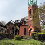
- Historic Astoria Church Damaged in Blaze, Neighbors Blame Teen Trespassers
- Ravenswood: Queens’ Own Beverly Hills

Social Media
Advertisement